Exploring Tumour-Specific Fluorescent Probes in Oesophagogastric Cancer
Authors
Michael Bozin1,2, Nicholas Clemons1, Laura Edgington-Mitchell2, Gavin Wright1, Wayne Phillips1 and Cuong Duong1
Affiliations
- Department of Cancer Surgery and Research, Peter MacCallum Cancer Centre, Melbourne, Australia
- Department of Pharmacology, Bio21 Research Institute, University of Melbourne, Australia
Background
Intra-operative molecular imaging (IMI) is an emerging field that utilises tumour-targeting fluorescent probes to improve oncological outcomes. Quenched activity-based probes (qABP), designed to measure enzyme activity in-vitro, have been repurposed to target tumour-expressed proteases known as cathepsins in-vivo. Fluorescence is initially suppressed by an inhibitory quencher until the qABP covalently binds to cathepsins and the quencher is released. This may improve the contrast between normal tissue and tumour. Our research group sought to obtain pre-clinical evidence for translating qABP for Oesophagogastric cancer surgery.
Methods
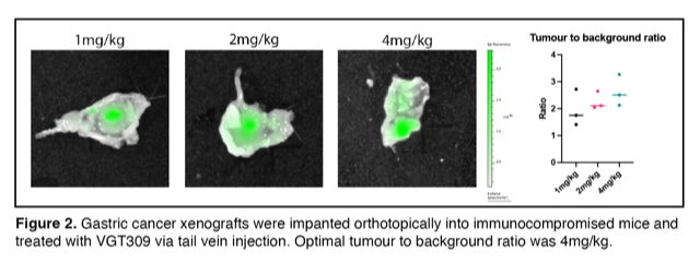
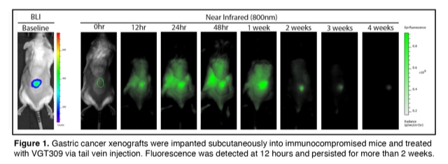
Cathepsin activity was assessed in Oesophagogastric cancer cell lines and patient biopsies with two qABPs, BMV109 and VGT309. Cell lines and tissue samples were analysed using in-gel fluorescence (Amersham Typhoon®) and Western blot. Cell line xenografts were established in immunocompromised mice using subcutaneous and orthotopic models. Tumour-bearing mice were injected with VGT-309 and imaged at specific timepoints using the IVIS® spectrum imager.
Results
Cathepsin activity was found in Oesophagogastric cell lines and selectivity of qABPs demonstrated with a cathepsin inhibitor. Matched biopsies were collected from patients prior to and after neoadjuvant chemotherapy. Baseline tumour biopsies exhibited significantly higher cathepsin activity than normal oesophageal and gastric biopsies (p <0.05). Oesophageal and gastric adenocarcinoma xenografts were identified in mice as early as 12 hours post injection of VGT-309, exhibiting a 2.5-fold increase in fluorescence compared to normal background stomach. Bio-distribution analysis demonstrated that VGT-309 accumulated in liver and kidney, but less so in the murine stomach.
Conclusion
Translating qABPs into the operating room has the potential to detect early cancer, reduce positive margin rates and identify lymph node metastasis. Our research group continues to investigate qABPs, including determining their sensitivity to metastasis, with the aim of translating this technology into a phase 1 clinical trial.